
Contents

All living organisms are made up of cells and new cells are produced when live cells divide. The cell is the smallest unit of life in an organism. The cell lives and, as a result, the organism lives. Whatever an organism does for survival it does for the survival of its cells.
Microscopes
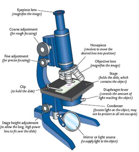
The first microscopes were composed of a single lens just like a magnifying glass. They were called simple microscopes. Later microscopes were designed using 2 lenses. They are called compound microscopes. The top lens through which you look is called the eyepiece while the lower lens that is close to the slide is called the objective lens. They are called compound microscopes. The magnification power of a compound microscope is calculated by multiplying the magnification of the 2 lenses. Since these microscope use light to see the objects they are called light microscopes.
Below is a diagram of a compound light microscope. Learn the various parts. You will be using the microscope in your biology study.
Diagram of a Microscope
Robert Hooke
In 1665 Robert Hooke used a light microscope to study cork. He noticed that the cork was composed of many small boxes. He named the compartment cells because they reminded him of prison cells.
It is now known that all living things are composed of cells. They are measured using the unit micrometer. The symbol for micrometer is m. A m is one thousandth of a millimetre.
Animal Cells
The diagram below is an animal as may be seen using a light microscope. All the living matter of a cell is called protoplasm. The cell is surrounded by a cell or plasma membrane. The nucleus is the control centre of the cell. The cytoplasm surrounds the nucleus. The cytoplasm is everything within the cell except for the nucleus. There are many small organelles within the cytoplasm. This is where most of the cell s activities take place. The cytoplasm is composed of 90% water. They cannot be seen using a light microscope. They will be discussed later.
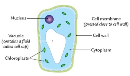
Plant Cells
The diagram below is a plant cell as may be seen using a light microscope. All the living matter of a plant cell is also called protoplasm. The cell is surrounded by a cell or plasma membrane. Unlike the animal cell the plant cell also has a cell wall surrounding it. This is made of cellulose and is very rigid. It supports the plant cell. The nucleus is the control centre of the cell. The cytoplasm surrounds the nucleus. The vacuole is a storage area for the plant cell. The vacuole contains cell sap. This is made of sugars, salts, and pigments. The chloroplasts contain chlorophyll. This is where photosynthesis occurs within the cell.
The Ultrastructure of Cells
With the invention of the electron microscope a whole new world was open up to scientists. Most light microscopes will enlarge a specimen up to 1000 times (1000X) but the electron microscope enlarge the specimen 250,000X and higher! Using these microscopes scientists were able to discover parts of the cell never seen or known of before. The fine detail of a cell when seen by an electron microscope is called ultrstructure.
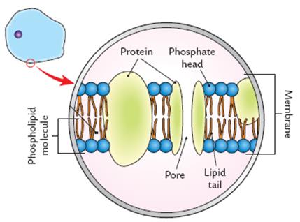
Cell Membrane (Plasma Membrane)
Below is a diagram of a part of the plasma membrane. Note that they are composed of phospholipid molecules and protein. The phosphate heads of each molecule is on the outside of the structure while the lipid tail of each are on the inside part of the membrane. The protein location varies along the membrane. Note that there are pores along the membrane. This is where materials enter and leave the cell. The parts of the membrane move around constantly. That is why the membrane is said to be fluid. This is called the fluid mosaic model of the plasma membrane.
Plasma Membrane Functions
The membranes allow some materials to enter the cell but not all materials. Thus, the membrane is said to be semipermeable. Water, oxygen and carbon dioxide freely pass through it, many other chemicals cannot. Another term used for this aspect of the membrane is selectively permeable.
The membrane keeps cell contents together allowing efficient coordination of its activity. It helps the cell keep its contents.
Give support to the cell.
Recognise (sense) molecules that touch them.
Nucleus
The nucleus is the control centre of the cell. It is surrounded by a nuclear membrane that allows molecules to enter and leave the nucleus similar to the plasma membrane.
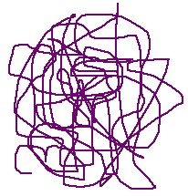
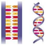
The nucleus contains DNA (deoxyribonucleic acid). The DNA is arranged in groups called chromosomes. This is the genetic material of the cell. Every organism has a specific number of chromosomes in each nucleus of each of its cells. Humans have 46 chromosomes in every cell while roundworms have 2. When not dividing the chromosomes are called chromatin. They become elongated and interwoven at this stage.
A molecule of DNA when the cell is not dividing (in chromatin form):
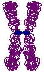
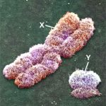
A chromosome as seen at the start of cell division:
Genes are located on the chromosomes. These are the structures that control the production of protein. In this way the genes determine the characteristics of the living thing.
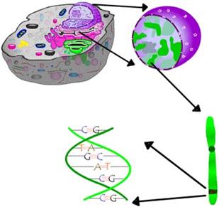
The nucleus of a cell contains chromosomes, on which are the genes consisting of the DNA that codes for proteins, the main ingredients of living beings.
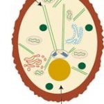
Nuclear Pores
Nuclear pores are openings through which materials enter and leave the nucleus. Large molecules can pass between the cytoplasm and the nucleus through these pores. 1 example is RNA from nucleus to cytoplasm and nucleotides from cytoplasm to nucleus. This will be discussed later in another chapter of your text.
![]()
The arrows show a nuclear pore.
Nucleolus
The nucleolus is the area where ribosomes are made. Ribosomes are made of RNA. The nucleolus contains a lot of rRNA (ribosomal RNA)
Mitochondria
The mitochondria are called the powerhouses of the cell. This is where cellular respiration occurs. The end product of cellular respiration is energy. Muscle and liver cells have many mitochondria and produce a lot of energy. The mitochondria have 2 membranes. The inner membrane is folded. Here, at the folds, is where the energy is releases. The more folds it has the more energy is released. Foldings increase during exercise and activity while they decrease during rest.
In general:
1. The aerobic steps of respiration occur here.
2. 36 of the 38 ATPs (energy molecules) from one molecule of glucose are produced in the mitochondrion.
3. Liver, muscle and nerve cells are rich in mitochondria.
4. Bone and fat cells have low numbers of mitochondria.
5. Root hair cells and meristematic cells of plants have large numbers of mitochondria.
6. Stem and root ground tissue cells of plants are low in mitochondria.
Chloroplasts
This is where photosynthesis takes place in green plants. The green pigment is called chlorophyll and is stored in the chloroplasts.
Below is the ultrastructure of a chloroplast.
Ribosomes
Ribosomes are composed of RNA and protein. They function in protein synthesis. They make protein using amino acids. This will be discussed in a later chapter of your text.
Other Cellular Structures
Below is a generalised ultrastructure of an animal and a plant cell. We have not discussed all the parts labelled since they are not part of your syllabus or will be discussed later in the syllabus. Below the diagrams are definitions to the terms not discussed.
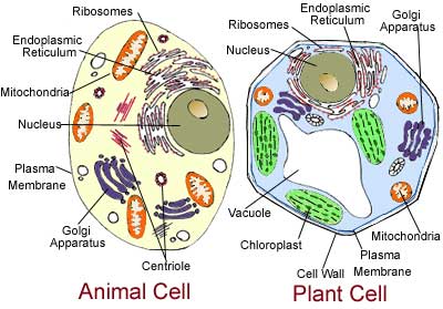
Golgi Apparatus – a flattened, layered, sac-like organelle that looks like a stack of pancakes and is located near the nucleus. It produces the membranes that surround the lysosomes. The Golgi body packages proteins and carbohydrates into membrane-bound vesicles for “export” from the cell.
Endoplasmic Reticulum- The endoplasmic reticulum contains a network of branching and joining tubules 400 to 700 angstroms in diameter (1 angstrom equals 10-9 m). It has been calculated that 1 ml of liver tissue contains about 11 square meters of endoplasmic reticulum. The encircling membranes are about 50 to 60 angstroms thick and have the same substructure as the plasma membrane. Two patterns are found in the cell, the smooth endoplasmic reticulum and the rough endoplasmic reticulum. The rough endoplasmic reticulum is covered by an evenly spaced arrangement of ribosomal granules. The smooth endoplasmic reticulum lacks ribosomes, which synthesize proteins. The smooth endoplasmic reticulum, rich in a wide variety of enzymes, is most common in cells that are involved in the synthesis of lipids, triglycerides, lipoprotein complexes, and steroids.
Centriole- Centrioles generally appear in animal cells as two cylinders at right angles to one another, close to the nucleus. When viewed with an electron microscope, the cylinders show up as nine bundles of tiny microtubules arranged in a circle. The centrioles help to form the spindle fibres. Spindle fibres are microtubules that move chromosomes around when the cell is dividing.
Differences Between a Plant and an Animal Cell
Below is a chart showing the general differences between a plant and an animal cell.
|
Plant cells |
Animal cells |
|
Possess rigid cell walls |
No cell walls |
|
Have green chloroplasts |
No chloroplasts |
|
Contain chlorophyll |
Do not contain chlorophyll |
|
Thin lining of cytoplasm |
Most of cell is cytoplasm |
|
Vacuole filled with cell sap |
Small (if any) vaculoes |
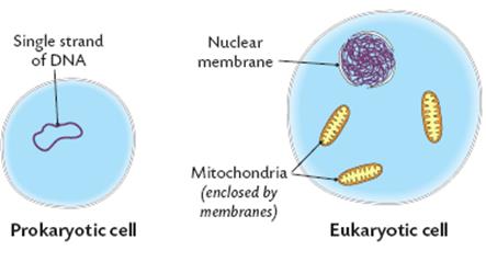
Prokaryotic and Eukaryotic Cells
All living things can be categorized as being prokaryotic or eukaryotic.
Prokaryotic cells have no nucleus. They do not have membrane-bound organelles such as nuclei, mitochondria or chloroplasts. All prokaryotes are placed in the Kingdom Monera i.e. the bacteria.
Eukaryotic cells have a membrane-bound nucleus. Membrane-bound organelles such as nuclei, mitochondria and chloroplasts are only present in eukaryotic cells. The Protista, Fungi, Plants and Animals are eukaryotic organisms.
We thank Americas Cardroom for supporting us. You can learn more about Americas Cardroom at https://gpsts.org/americas-cardroom-bonus-code-review/
Submit your review | |