Contents
The process of excretion eliminates metabolic wastes from our bodies. It also helps us maintain a constant internal body temperature.
The Skin
The skin plays a large role in the elimination of wastes and the regulating of body temperature.
The Structure of the Skin
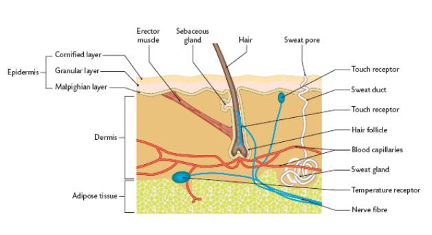
The skin has two layers. There is the outer epidermis and the inner dermis layers. The adipose layer is a layer below the dermis, which contains fat-rich cells.
The Epidermis
The malphigian layer is where cell division takes place to produce new epidermal cells. As these cells move out toward the surface they produce a protein called keratin. This protein causes the cells to become hardened. There are no capillaries in the outer layers of the skin so the cells die. These cells continually wear away.
Also in the malphigian layer are cells that produce melanin. This is a pigment, which varies in colour from light brown to black in different people. This pigment gives our hair, eyes, and skins their colour. Melanin also protects the skin from harmful ultraviolet rays from the sun. Its production increases when the skin is exposed to the sun’s rays. This causes people to tan.
The Dermis
The dermis is live connective tissue. It contains collagen which helps strengthen the skin. It also contains other structures seen on the diagram and discussed later.
Functions of the Skin
The skin:
Protects against physical injury.
Sebum, produced in the sebaceous glands, keeps the hair moist and flexible and prevents the drying out of the skin
Provides some protection for the body against many pathogenic microbes and chemical agents.
Fat in the andipose layer acts as a food store.
Helps prevent excessive water absorption by imparting water resistance to the skin.
Is the sensory organ for temperature, pressure, touch and pain.
Melanin protects underlying tissues from UV light.
Plays a role in metabolism, including vitamin D synthesis following the exposure to ultraviolet light. Lack of vitamin D leads to soft bones that may bend under the weight of the body and may result in bowed legs, called rickets. Vitamin D facilitates the absorption of calcium and phosphorus from the small intestine.
Acts as an agent of excretion. Sweat contains water and salts. Sweat glands pass these out or the sweat pores.
Plays a major part in temperature regulation of the body.
Temperature Regulation
In cold conditions:
1. Goose bumps also called goose pimples, goose flesh chicken skin or cutis anserina, are the bumps on a person’s skin at the base of body hairs which involuntarily develop when a person is cold or experiences strong emotions like fear. The reflex of producing goose pimples is known piloerection. It occurs not only in humans but also in many other mammals; a prominent example is porcupines, which raise their quills when threatened.
Goose bumps are created when tiny muscles at the base of each hair, known as erector muscles contract and pull the hair erect. The reflex is started by the sympathetic nervous system, which is in general responsible for many fight-or-flight responses.
Goose bumps are often a response to cold: in animals covered with fur or hair, the erect hairs trap air to create a layer of insulation. Goose bumps can also be a response to fear: the erect hairs make the animal appear larger, in order to intimidate enemies. This can for example be observed in frightened cats. In humans, it can even extend to piloerection as a reaction to hearing nails scratch on a chalkboard.
2. Blood vessels in the skin contract (get narrower) so as to reduce heat loss from the blood near the surface of the skin.
3. Shivering is a bodily function in response to cold in warm-blooded animals. When the core temperature of the body drops, the shivering reflex is triggered. Muscle groups around the vital organs begin to shake in small movements in an attempt to create warmth by expending energy. Located in the hypothalamus of the brain is an area called the primary motor center for shivering. This area is normally inhibited by signals from the heat center of the brain but is excited by cold signals from the skin and spinal cord. Therefore, this center becomes activated when the body temperature falls even a fraction of a degree below a critical temperature level.
4. Fat in the andipose layer insulates our bodies. Many animals (ducks, polar bears, whales, seals) produce large amounts of insulating fat.
In Warm Conditions:
1. Sweat is produced and released in warm conditions. The evaporation of the sweat cools our bodies as the heat is released from the sweat.
View an animation of the action of sweat glands
2. Blood vessels in our skin dilate (get wider). As more blood comes to the surface of the skin more heat from the blood is released.
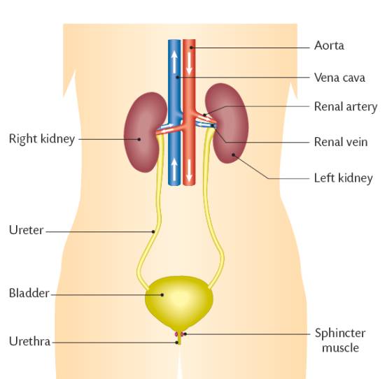
The Urinary System
The urinary system is diagramed below. It is the main system through which our bodies maintain homeostasis. Through this system the salt and water balance of our bodies is maintained as well as the concentration of body fluids and the elimination of metabolic wastes.
The Kidneys
(A General Discussion for Ordinary Level)
The kidneys are the main excretory organ of the body. They excrete water, salts, and urea in the form of urine.
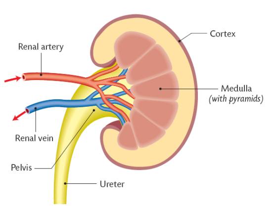
The blood from the aorta enters the kidneys through the renal arteries. This blood is rich in waste products it collected from the cells of the body. Every minute about 20% of our blood enters the kidneys. The kidneys are about the size a fist, and lie in the back. They are partly protected by the lower ribs. Each kidney contains about a million filters. Each is called a glomerulus and are located in the outer cortex of the kidneys. As a result of filtration small substances are taken out of the bloodstream and put into the kidneys. Some of the useful materials are then reabsorbed into the blood.
Unwanted wastes and toxic products are left in the kidneys and are then formed into a liquid called urine. The formation of urine occurs in millions of tiny kidney tubules called nephrons. Also, hydrogen ions as well as potassium are secreted from the blood into the kidneys.
The urine flows from the medulla into the pelvis of each kidney. It is then carried by the ureters to the bladder.
The purified blood leaves the kidneys through the renal veins and then travels to the vena cava.
The Bladder
An adult bladder can hold about 800 ml. of urine. Two sphincter muscles are located where the bladder and the urethra meet. These muscles open automatically in babies when the bladder is ½ full. Adults learn to control these muscles. The urethra emerges through the penis in males and close to the vagina in females.
To review: The main functions of the kidneys are:
1. Excretion of water, salts and urea. (urine)
2. Control of water content of the blood and body fluids. Varying the amount of urine produced does this. On hot days less urine is produced than on cold days.
3. Control of salt concentration of the blood and body fluids. This is necessary n order for our body cells to have the same salt concentration as our blood. If this did not happen then the osmosis or movement of water between our body cells and our blood would be hindered.
4. Control of the pH of the blood and body fluids. This is done by varying the acidity of our urine in order to maintain a blood pH of 7.4
The Nephron
Blood is pumped from the heart through large blood vessels to the kidneys. The kidneys are about the size a fist, and lie in the back. They are partly protected by the lower ribs. Each kidney contains about a million filters. Each is called a glomerulus.

Inside the kidney the blood vessels continue to divide until they become so small that they can no longer hold water. These tiny vessels are known as capillaries. The glomerulus is the filtering unit and contains the leaky capillary. The filtrate collects into a sac called Bowmans capsule and drains into the proximal tubule.
The tiny capillaries filter minerals, wastes and water but retain red cells, proteins and large molecules. This process is known as filtration. The proteins that are not filtered remain in the capillary and create an oncotic pressure due to osmosis. Filtration depends upon the surface area of the filter and the permeability of the membrane that surrounds the capillary. It also depends upon the systemic pressure and its counter, the pressure caused by osmosis.
The filtered blood is returned to the body through the veins. The fluid and its substances must now travel through a long and winding tubular pathway, first through the cortex, or outer portion of the kidney, then the medulla, the deep portion. There the tubule makes a sharp hairpin turn and travels back up to the cortex and its parent glomerulus. After looping through the cortex it connects to a collecting tubule and makes its final descent. The nephron is the combination of the filtering unit (glomerulus), its tubule and collecting duct. Together, they filter the blood, process the filtrate and make urine.