Contents
The nervous system allows the animal to quickly detect, communicate and co-ordinate information about its external and internal environment so it can make efficient appropriate responses for survival and/or reproduction.
The two major parts of our nervous system are the central nervous system (CNS) and peripheral nervous system (PNS).
The CNS is made of the brain and spinal cord.
The cranial nerves, spinal nerves and ganglia make up the PNS. The cranial nerves connect to the brain. The cranial and spinal nerves contain the axons (fibres) of sensory and motor nerve cells.
Nerve cells areas are also known as neurons. Neurons are the basic unit of the nervous system. They carry information or impulses as electrical signals from one place to another in the body.
Types of Neurons
There are 3 types of neurons:
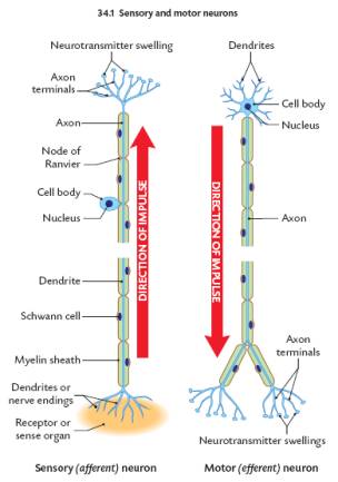
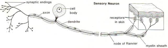
Sensory Neurons- Sensory neurons carry electrical signals (impulses) from receptors or sense organs to the CNS. Sensory neurons are also called afferent neurons. The cell body of sensory neurons is outside the CNS in ganglia.
Motor Neurons- Motor neurons carry impulses from the CNS to effector organs Motor neurons are also called efferent neurons. The cell bodies of motor neurons are inside the CNS.
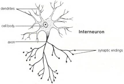
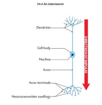
Interneurons- These are also called intermediate, relay, or associative neurons. They carry information between sensory and motor neurons. They are found in the CNS.
The Structure of Neurons
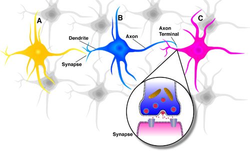
A Neuron consists of THREE MAIN PARTS:
A. CELL BODY – The largest part, contains the nucleus and much of the cytoplasm (area between the nucleus and the cell membrane), most of the metabolic activity of the cell, including the generation of ATP (Adenine Triphosphate Compound that Stores Energy) and synthesis of protein.
B. DENDRITES – Short branch extensions spreading out from the cell body. Dendrites Receive STIMULUS (Action Potentials) and carry IMPULSES from the ENVIRONMENT or from other NEURONS AND CARRY THEM TOWARD THE CELL BODY.
C. AXON – A Long Fibre that CARRIES IMPULSES AWAY FROM THE CELL BODY. Each neuron has only ONE AXON. The Axon Ends in a series of small swellings called AXON TERMINALS.
Neurons may have Dozens or even Hundreds of DENDRITES but usually ONLY ONE AXON.
Sensory Neuron or Afferent Neuron: Moving away from a central organ or point. Relays messages from receptors to the brain or spinal cord.
Motor Neuron or Efferent Neuron – Moving toward a central organ or point. Relays messages from the brain or spinal cord to the muscles and organs.

Interneurons- Relay message from sensory neurone to motor neurone. Make up the brain and spinal cord.
|
|
Sensory neuron |
Interneuron |
Motor Neuron |
|
Length of Fibers |
Long dendrites |
Short |
Short |
|
Location |
Cell body and |
Entirely |
Dendrites and |
|
Function |
Conduct |
Interconnect |
Conduct |
The Axons of most Neurons are covered with a Lipid Layer known as the MYELIN SHEATH. The Myelin Sheath both Insulates and Speeds Up transmission of Action Potentials through the Axon. In the Peripheral Nervous System, Myelin is produced by SCHWANN CELLS, which surround the Axon. GAPS (NODES) in the Myelin Sheath along the length of the Axon are known as the NODES OF RANVIER. These gaps allow the impulses to travel faster than if they travelled along the entire length of the neuron.
The Axon ends with many small swellings called AXON TERMINALS. At these Terminals the neuron may make contact with the DENDRITES of another neuron, with a RECEPTOR, or with an EFFECTOR. RECEPTORS are special SENSORY NEURONS in SENSE ORGANS that RECEIVE Stimuli from the EXTERNAL ENVIRONMENT. EFFECTORS are MUSCLES or GLANDS that bring about a COORDINATE RESPONSE.
The Synapse
The points of contact at which impulses are passed from one cell to another are known as THE SYNAPTIC CLEFT OR SYNAPSE. Neurons that transmit impulses to other neurons DO NOT actually touch one another. The Small Gap or Space between the axon of one neuron and the dendrites or cell body on the next neuron is called the Synapse. One importance of the presence of Synapses is that they ensure one-way transmission of impulses in a living person. A nerve impulse CANNOT go backward across a Synapse.
The Axon Terminals at a Synapse contain tiny vesicles, or sacs called neurotransmitter swellings. These tiny swellings are filled with CHEMICALS known as NEUROTRANSMITTERS. Acetylcholine (Ach) and noradrenialin, also called norepinephrine, are 2 of the main neurotransmitters.
A NEUROTRANSMITTER is a chemical substance that is used by one neuron to signal another. Some are made in the cell body while others are made in the neurotransmitter swellings. The impulse is changed from and Electrical Impulse to a Chemical Impulse (Electrochemical Impulses). The molecules of the neurotransmitter diffuse across the gap and attach themselves to SPECIAL RECEPTORS on the membrane of the neuron receiving the impulse. This now causes the electrical impulse to be regenerated. After the neurotransmitter relays it message it is rapidly REMOVED or DESTROYED, thus halting its effect. ENZYMES, taken up again by the axon terminal and recycled, may break down the molecules of the neurotransmitter or they may simply diffuse away.
Synapses are the slowest part of the nervous system. The advantage to having many neurons, with gaps between them, is that we can control and receive information from different parts of the body at different times. They also ensure one-way transmission of impulses in a living person. The number of synapses associated with each neuron varies from 1000 for a cell body of the spinal cord to up to 10,000 for cell bodies in the brain.
To Review: The main functions of the synapse are:
1. To transmit impulses from one neuron to another neuron or to an effector.
2. To control the direction of the impulse. Impulses can only go one way. The neurotransmitter swellings are only found on the presynaptic side of the synapse. Thus, the impulse can only travel from the presynaptic side to the postsynaptic side.
3. To prevent over stimulation of effectors. Constant stimulation causes neurotransmitter production to cease. In this way we get used to stimuli such as pain or noise.
4. Certain chemicals can block the impulse. This is why doctors prescribe certain drugs for pain relief.
Nerve Impulses
Nerve impulses are electrical as they run along the nerve. They then become chemical as the travel over the synaptic cleft.
When a neuron receives a stimulus of sufficient strength the electrical current moves along the dendrite and axon to the neurotransmitter swellings. The movement of ions causes these electrical impulses.
Resting Neuron
When a neuron is not carrying an impulse the inside of the axon has a negative charge and the outside has a positive charge.
The threshold is the minimum stimulus needed to cause an impulse to be carried. It must be of sufficient strength. Not all stimuli cause an impulse. A stimulus below the threshold has no effect on the neuron. Some people have higher thresholds for pain, heat or other stimuli. This means they can tolerate a stronger stimulus before their nervous system reacts with an impulse.
All Or Nothing Law
The “All or Nothing” Law states that if the threshold is reached an impulse is carried, but if the threshold is not reached then there will be no impulse. It doesn’t matter how strong the stimulus. The same impulse is sent regardless of strength. The sensitivity to mild or severe pain depends on the number of neurons stimulated as well as the frequency of their stimulation.
Movement of the Impulse
When the threshold is reached the axon or dendrite changes. The inside, at the point of the stimulation, becomes positive and the outside becomes negative. This creates unlike charges along the length of the neuron and the impulse travels along the neuron. This is called the action potential. Once the impulse moves along, the area behind the impulse is changed back to its normal negative (resting) state.
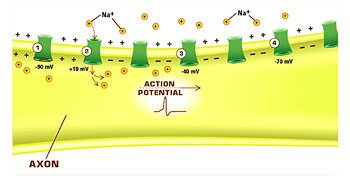
Below is a cross-section of an axon, with an action potential (AP) moving from left to right. The AP has not yet reached point 4; the membrane there is still at rest. At point 3, positive sodium ions are moving in from the adjacent region, depolarising the region; the sodium channels are about to open. Point 2 is at the peak of the AP; the sodium channels are open and ions are flowing into the axon. The AP has passed by point 1; the sodium channels are inactivated, and the membrane is hyperpolarized.
Refractory Period – While the ions are moving in and out of each region of the neuron, there is a brief period during which the neuron is unable to have another action potential. This delay is called the refractory period.
In Summary:
The resting potential tells about what happens when a neuron is at rest. An action potential occurs when a neuron sends information down an axon, away from the cell body. Neuroscientists use other words, such as a “spike” or an “impulse” for the action potential. The action potential is an explosion of electrical activity that is created by a depolarising current. This means that some event (a stimulus) causes the resting potential to move toward 0 mV. When the depolarisation reaches about -55 mV a neuron will fire an action potential. This is the threshold. If the neuron does not reach this critical threshold level, then no action potential will fire. Also, when the threshold level is reached, an action potential of a fixed sized will always fire…for any given neuron, the size of the action potential is always the same. There are no big or small action potentials in one nerve cell – all action potentials are the same size. Therefore, the neuron either does not reach the threshold or a full action potential is fired – this is the “ALL OR NONE” principle.
Neural Impulse Terms
A. Neural impulse – takes the same path all the time – it is a process of conducting information from a stimulus by the dendrite of one neuron and carrying it through the axon and on to the next neuron. Let’s take a look at what’s involved in the neural impulse:
1) ions – we have positively (+) and negatively (-) charged particles called ions. For the neural impulse, however, we are only concerned with Sodium (Na+) and Potassium (K+).
2) selectively permeable membrane – the outer membrane of the neuron is not impermeable, but instead selectively allows some ions to pass back and forth. The way it selects is easy – it has pores that are only so big. So, only very small ions can fit through. Any large ions simply can’t pass through the small pores.
3) charge of the neuron – inside the neuron, the ions are mostly negatively charged. Outside the neuron, the ions are mostly positively charged. In this state (with mostly negative charge inside and positive charge on the outside) the neuron is said to be Polarized.
4) resting potential – while the neuron is Polarized, it is in a stable, negatively charged, inactive state The charge is approx. -70 millivolts, and it means that the neuron is ready to fire (receive and send information).
5) stimulus – eventually, some stimulation occurs (ex. hand to close to a flame), and the information is brought into the body by a sensory receptor and brought to the dendrites of a neuron.
6) action potential – once the stimulation (the heat) reaches a certain threshold (come to later) the neural membrane opens at one area and allows the positively charged ions to rush in and the negative ions to rush out. The charge inside the neuron then rises to approx. +40 mv. This only occurs for a brief moment, but it is enough to create a domino effect.
7) repolarization – the neuron tries to quickly restore its charge by pumping out the positively charged ions and bringing back the negative ones. This can occur fast enough to allow up to 1,000 action potentials per second.
8) absolute refractory period – after the action potential occurs, there is a brief period during which the neuron is unable to have another action potential. Then the charge inside the neuron drops to about -90 mv (refractory period) before restoring itself to normal.
9) speed of an action potential – can travel from 10-120 meters/sec. The speed depends on whether a myelin sheath is present or not. If there is no myelin sheath then the impulse travels all along the axon or dendrite. This acts to slow down the impulse. If there is a myelin sheath then the impulse charges can only move in and out at the nodes of Ranvier. These impulses move more rapidly than the non-myelinated neurons. Also, the larger the diameter of the axon or dendrite the faster the impulse.
10) all-or-none law – a neural impulse will either occur or not. There is no in between. Once the threshold is reached, there is no going back, the neural impulse will begin and will go through the complete cycle.
11) Threshold – a dividing line that determines if a stimulus is strong enough to warrant action. If the threshold is reached, an action potential will occur.
The Central Nervous System
The Brain
After sensory neurons carry impulses most eventually reach the brain. The brain acts to interpret, sort, and process the incoming impulses and then decide on a response.
The brain s grey matter is composed of cell bodies and synapses. The white matter is made of nerve fibres (axons and dendrites). There are about 12,000 million neurons that form the brain.
3 membranes called the meninges protect the brain and the spinal cord. The space between the inner 2 membranes is filled with a liquid called cerebrospinal fluid. There is a total of about 100 mL of this liquid in the CNS. It protects the CNS by acting as a shock absorber.
Inflammation of the meninges causes a sometimes-serious condition called meningitis. Refer to your text for a description of viral and bacterial meningitis.
Structure of the Brain:
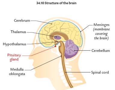
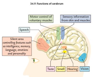
The Cerebrum
1. Largest part of the brain
2. Contains about 75% of the total neurons of the brain
3. Divided into 2 halves: The right and left cerebral hemispheres
4. Control:
a. voluntary movements
b. receiving and interpreting impulses from sense organs
c. thinking
d. intelligence
e. memory
f. language
g. emotions
h. judgement
i. personality
The right hemisphere controls the left side of the body while the left hemisphere controls the right side of the body.
Each hemisphere is specialised for different functions.
Generally:
The left side is dominant for: The right side is dominant for:
1. hand use 1. art
2. language 2. music
3. mathematics 3. shape recognition
4. analysis 4. emotional responses
5. logic
The outer part of the cerebrum is grey and called the cerebral cortex. It is divided into 4 lobes. Each lobe controls specific functions:
Notice that there are many infolds of the cerebral cortex. This gives it a larger surface area. This allows for more interconnections between different parts of the brain and for more efficiency.
The inner part of the cerebrum is white matter. It is made of millions of nerve fibres. These nerve fibres connect different areas of the cerebral cortex as well as the 2 sides of the brain.
The Cerebellum
1. Second largest part of the brain
2. Heavily folded
3. Controls muscular coordination
4. Allows for smooth, refined muscular action
5. Responses involuntary once they are learned
The Medulla Oblongata
1. Connects the brain with the spinal cord
2. Contains clusters of nerve cells that control involuntary actions such as:
a. breathing
b. blood pressure
c. swallowing
d. coughing
e. salivation
f. sneezing
g. vomiting
The Thalamus
1. Located below the cerebrum
2. Acts as a sorting centre for the brain. It relays incoming impulses to the relevant part of the brain.
The Hypothalamus
1. Lies below the thalamus
2. Regulates the internal environment (homeostasis) of the body by monitoring:
a. blood temperature
b. appetite
c. thirst
d. osmoregulation
e. blood pressure
3. Regulates the production of many hormones of the pituitary gland.
The Spinal Cord
The spinal cord is a long, fragile tubelike structure that begins at the end of the brain stem and continues down almost to the bottom of the spine (spinal column). The spinal cord consists of nerves that carry both incoming and outgoing messages between the brain and the rest of the body. It is also the centre for reflexes, such as the knee jerk reflex. Like the brain, the spinal cord is covered by three layers of tissue called meninges. The spinal cord and meninges are contained in the spinal canal, which runs through the centre of the spine. In most adults, the spine is composed of 26 vertebrae, which are the individual bones of the back. Just as the skull protects the brain, vertebrae protect the spinal cord. The vertebrae are separated by disks made of cartilage, which act as cushions, reducing the forces generated by movements such as walking and jumping.
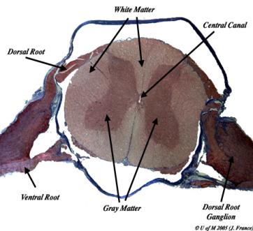
Like the brain, the spinal cord consists of grey and white matter. The butterfly-shaped centre of the cord consists of grey matter. The grey matter contains dendrites and cell bodies. The front or ventral root contain motor nerves, which transmit information from the brain or spinal cord to muscles, stimulating movement. The back or dorsal root contain sensory nerves, which transmit sensory information from other parts of the body through the spinal cord to the brain. The surrounding white matter contains columns of axons that carry sensory information to the brain from the rest of the body (ascending tracts) and columns that carry impulses from the brain to the muscles (descending tracts). There are a total of 31 pairs of spinal nerves. These carry impulses to and from the spinal cord.
Reflex Action
A reflex is the simplest, quickest form of activity in the nervous system. It is an automatic, involuntary, unthinking response to a stimulus. The reflex arc are the neurons that form the pathway of the impulses of a reflex. Examples of reflex actions are breathing, eye blinking, iris size, and many protective actions such as moving away from a burning flame. (see below)

When we move our finger away from a flame we are performing a withdrawl reflex. These satge of this reflex are as follows:
1. The finger is the receptor. It contains sensory neurons.
2. Sensory neurons carry the impulse to the sensory nerves in the dorsal root.
3. An interneuron carries the impulse across the spinal cord to the motor neurons in the ventral root. At the same time, another neuron takes the impulse to the brain.
4. The motor neurons take the impulse to the effector (muscle) and the finger is pulled away. At the same time, the impulse reaches the brain and we are aware of the pain.
Another reflex action is The Knee Jerk Reflex:
The knee jerk reflex is one that you may have had tested at a check up at the doctor’s office. In this test, the doctor hits your knee at a spot just below your kneecap and your leg kicks out. Try it! Have a partner sit with his or her legs crossed so that his leg can swing freely. Hit his leg just below the knee with the side of your hand. DO NOT USE A HAMMER!!!! The leg will kick out immediately (if you hit the right place). The knee jerk reflex is called a monosynaptic reflex because there is only one synapse in the circuit needed to complete the reflex. It only takes about 50 milliseconds between the tap and the start of the leg kick. That is fast! The tap below the knee causes the thigh muscle to stretch. Information is then sent to the spinal cord. After one synapse in the ventral horn of the spinal cord, the information is sent back out to the muscle…and there you have the reflex.
Sponsors
We thank https://williamspromocodes.co.uk for supporting LeavingBio.net.